

Which Microbe Is The Smallest
Meet all five of the major groups of microbes, and find out what function they play along with their structure.
Microbes are organisms that are too small to exist seen with the unaided eye. They evolved long before the first plants and animals appeared and affect our lives in more means than we might wait.
Microbiologists are discovering new species of microbe at a faster rate than ever, thanks to advances in Deoxyribonucleic acid sequencing techniques. Current estimates suggest there could exist at least 1 billion different species of microbe on World, possibly more than. Microbial diversity is truly staggering, yet all these microbes can be grouped into 5 major types: Viruses, Bacteria, Archaea, Fungi, and Protists.
Let's look at each one in more detail.
Viruses
Viruses are the smallest of all the microbes. Their genome is made of either DNA or RNA (non both), and this is packaged inside a protein shell called a capsid. They are not fabricated of cells (acellular), cannot make their ain proteins and don't abound. Instead, they must infect a host cell and hijack its machinery to assemble new viruses. Viruses are usually only able to infect a express number of species living organism. Bacteriophages (Effigy 1) are viruses that infect bacteria. In some countries they are used to treat bacterial infections and there is renewed interest in phage therapy due to the alarming increase in antimicrobial resistant infections.
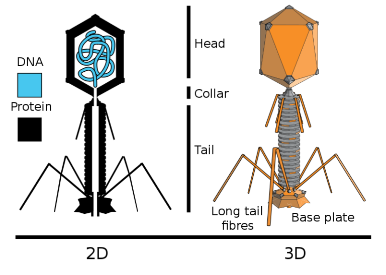
Figure 1: Structure of bacteriophage T2. The 2nd diagram on the left shows the structure of the protein capsid (black) and the nucleic acid genome (blue) packaged inside the head that is fastened (via the collar) to a hollow contractile tail. The 3D diagram on the right shows the caput has an icosahedral structure and the long tail fibres extend from the base plate at the bottom © Adenosine (original); en:User:Pbroks13 (redraw) CC Past-SA 2.5
Some viruses have a lipid envelope which they steal from membranes in the host prison cell. An example of an enveloped virus that you will be familiar with from News articles (and likely volition accept experienced the furnishings of personally) is influenza virus (Figure 2) which causes influenza or the 'flu'. The 1918-19 flu pandemic was the worst outbreak on record, infecting i third of the global population and killing around 50 million people. Some other instance is SARS-CoV-2, responsible for the current Covid-19 pandemic.
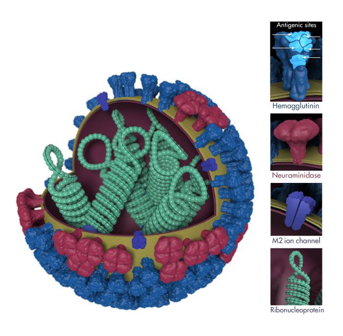
Figure 2: Construction of influenza virus. The eight segments of the RNA genome are packaged inside the virus particle, each piece of RNA is leap by protein that together course ribonucleoproteins (green). These are packaged inside the viral envelope (which the virus steals from the host cell membrane) that is embedded with viral proteins (light blue, red and dark blue) that play important roles in the virus replication bicycle © CDC/ Douglas Jordan; Dr. Ruben Donis, Dr. James Stevens Dr. Jerry Tokars, Flu Division
Whilst a few viruses can respond to changes in their environment, and you could argue that this is a grade of sensitivity (the S in MRS GREN – see below), they aren't capable of the majority of life processes. Almost scientists agree that viruses should therefore be considered not-living.
- K: Movement – the ability to change location
- R: Respiration – making energy from nutrients
- S: Sensitivity – responding to changes in the environment
- G: Growth – increasing in size
- R: Reproduction/replication – making new organisms
- East: Excretion – getting rid of waste product
- North: Diet – getting nutrients from the surround
In dissimilarity to viruses, bacteria, archaea, protists and fungi are all cellular, living organisms. They accept a DNA genome and make machinery to produce their own proteins. They are divided into two main groups based on their prison cell structure: Prokaryotes and Eukaryotes.
Prokaryotes
Leaner and Archaea are prokaryotes ("pro" = before, "karyote" = nucleus). They are single cells (unicellular) with a circular Dna genome that floats around in the cytoplasm. Many prokaryotes have one or more smaller circles of DNA called plasmids that carry additional genes. Bacteria and archaea have a very similar cell structure (Effigy 3). They practise not have a nucleus or membrane-bound organelles. Some prokaryotes have tail-like structures called flagella, which they use to swim through liquid, or fimbriae which enable them to stick to surfaces.
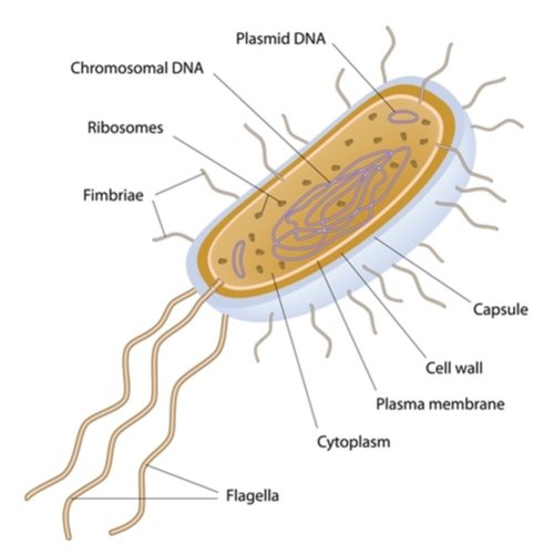
Figure 3: Labelled diagram of a bacterium showing the chief features of a prokaryotic cell
Bacteria
Bacterial cells are mostly circular (coccus, plural cocci) or rod-shaped (bacillus, plural bacilli), but a few have screw or corkscrew shapes. Some other defining features is the utilise of peptidoglycan as a component of their cell walls. Y'all almost certainly have millions of Escherichia coli leaner (Effigy 4) in your lower intestine. About strains of Due east. coli are harmless, but a few tin can cause serious food poisoning that tin can be fatal.
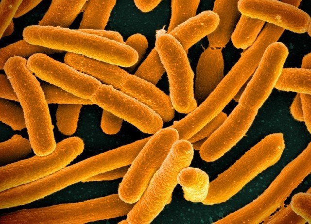
Figure 4: Scanning electron microscope (SEM) prototype of Escherichia coli © NIAID CC BY two.0
Archaea
Archaea wait similar in size and construction to bacteria but come in a wider diverseness of cell shapes. Many archaea live in hostile environments, some interact with other organisms, merely no archaea are known to cause disease. Different bacteria, they do not accept peptidoglycan in their cell walls. Many archaea live in farthermost environments, including Haloquadratum (Effigy five) which has ultra-thin (0.25 u (mu)m), square-shaped cells. Information technology is found all over the world in table salt lakes that are upwards to ten times saltier than bounding main water due to evaporation; so salty in fact, information technology would kill you if you were to drink it!
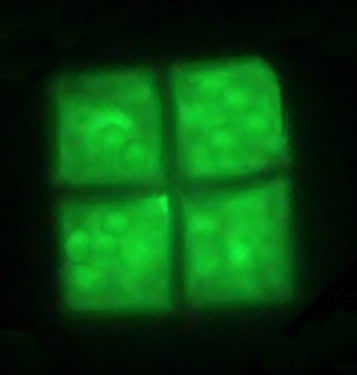
Figure v: Haloquadratum walsbyi, a salt-loving (halophillic) species of archaea © Rotational [Public domain]
Eukaryotes
The eukaryotes include protists, fungi, plants and animals. The defining feature of a eukaryotic cell is that information technology has a nucleus ("eu" = true, "karyote" = kernel or nucleus), in which the linear DNA genome is packaged into one or multiple chromosomes. Eukaryotic cells likewise contain multiple membrane-bound organelles, including mitochondria, that are not found in prokaryotic cells (Figure 6).
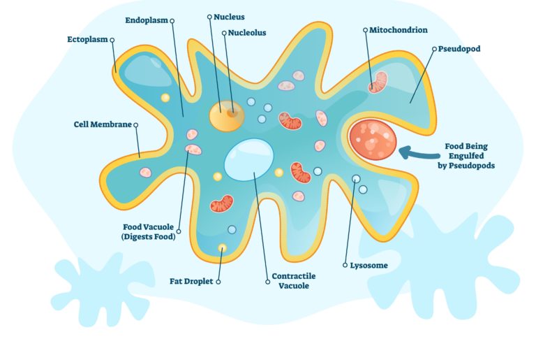
Figure vi: Labelled diagram of an amoeba to bear witness the main features of a eukaryotic jail cell. Note: this diagram lacks a label for ribosomes and Golgi apparatus – yous tin see these in the 3D model in a afterward Stride)
Protists
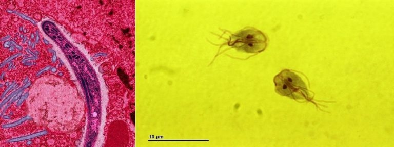
The protists are a very varied grouping consisting of all the eukaryotes that are not fungi, plants or animals. Most are unicellular and include plant-like algae, animal-like protozoa and fungi-like slime molds. Plasmodium (Figure vii: left) is a protist that causes the affliction malaria, responsible for the death of thousands of people every yr. Giardia (Figure 7: correct) is a protist that causes gastroenteritis (infection of the intestines) in humans. It has two nuclei – they make the cells wait like they have a face!
Click to expand
Figure 7: Left: Digitally colourised manual electron microscope (TEM) image of a Plasmodium sporozoite inside a mosquito gut cell © epitome by Ute Frevert; false colour by Margaret Shear CC BY 2.five. Correct: Calorie-free microscope image of Giardia intestinalis © Dr.. RNDr. Josef Reischig, CSc. (Author's annal) CC By-SA 3.0
Fungi
Fungi exist as either single cells (yeasts) or as multicellular organisms formed of sparse, branching tubular structures called hyphae. Some fungi are able to switch between these two forms (they are dimorphic) in response to environmental conditions eg temperature (Effigy 8).
One of the defining features of fungi is that their jail cell walls contain chitin. Arthropods such as crustaceans (crabs, lobsters, shrimp) and insects use chitin to form their exoskeleton – think of how tough a beetle is and you lot tin can encounter how this polymer provides structural support to fungal cells.
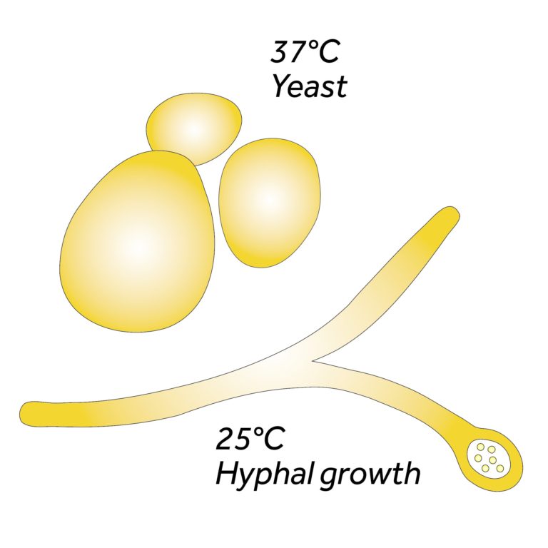
Figure 8: Some species of fungi that cause beast diseases are referred to as dimorphic (Greek di- twice, morphe – grade) because they grow as hyphae in ambient temperatures merely switch to yeast-like growth when causing an infection © Academy of Reading
Candida albicans (Figure 9) is a fungus that lives in the intestine and mouth of 50% of the human population. It ordinarily causes no damage, but if the normal community of microbes is disturbed (eg by taking antibiotics) or the immune arrangement is not functioning properly (eg in AIDS patients) it can crusade illness.
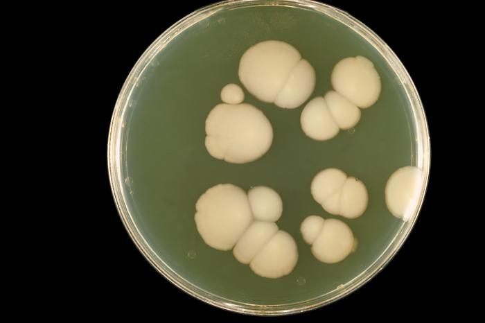
Figure ix: Colonies of the fungus Candida albicans growing on an agar plate © CDC/ Dr. William Kaplan
Recommended reading
- You tin find a summary of the key differences between the five major groups of microbe in this additional resource.

Which Microbe Is The Smallest,
Source: https://www.futurelearn.com/info/courses/introduction-to-microbiology/0/steps/51408
Posted by: gardnerstione.blogspot.com
0 Response to "Which Microbe Is The Smallest"
Post a Comment